JAAD study probes direct immunofluorescence in oral, esophageal lichen planus
Updated
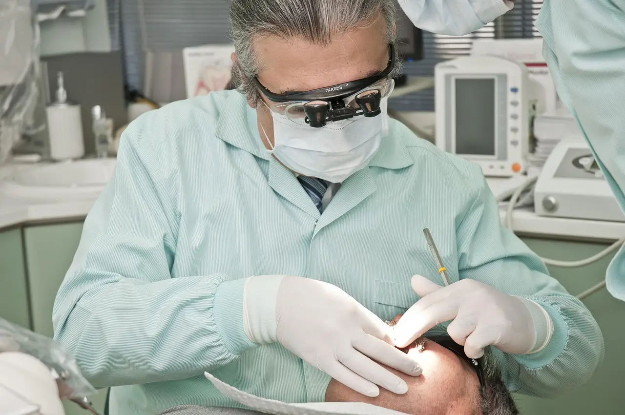
A study published in the Journal of the American Academy of Dermatology, Volume 95, Issue 5, dated November 2026, examines how often traditional direct immunofluorescent (DIF) findings appear in oral and esophageal lichen planus — and how sensitive those findings really are.
The authors — Justin Lyon, Annabelle Huntsman, Rachel Seifert, Charles Teames, David West, John Fang, John J. Zone, Christopher Hull, and Zachary H. Hopkins — set out to measure the prevalence and sensitivity of the classic DIF patterns clinicians have long used to support a lichen planus diagnosis on mucosal surfaces.
Why does this matter for dermatologists?
Oral lichen planus carries a small but real risk of malignant transformation, and esophageal disease can complicate swallowing in patients who often go undiagnosed for years. Histopathology alone frequently can't separate lichen planus from other lichenoid reactions, including drug eruptions and contact reactions to dental materials. DIF has served as the adjunct test, but its performance on mucosal tissue — rather than cutaneous lesions — has remained poorly characterized.
The Utah-based team, which spans dermatology and gastroenterology through the inclusion of John Fang, an esophageal specialist, anchored the work in biopsy-confirmed mucosal disease. John J. Zone and Christopher Hull bring long-standing expertise in immunodermatology and mucosal disease to the analysis.
What did the study measure?
The investigation focused on two endpoints stated in its title: the prevalence of traditional DIF findings in oral and esophageal lichen planus specimens, and the calculated sensitivity of those findings for the diagnosis. Traditional DIF criteria for lichen planus include cytoid bodies and fibrin deposition along the basement membrane zone, patterns originally validated in cutaneous disease.
By reporting prevalence and sensitivity together, the authors give clinicians a practical read on what a negative DIF result does — and doesn't — rule out when mucosal lichen planus is suspected.
What's the clinical takeaway?
The study lands in a diagnostic gap that matters for practice. Mucosal lichen planus often requires clinico-pathologic correlation, and overreliance on DIF positivity can delay diagnosis in patients whose biopsies lack the classic pattern. A sensitivity figure gives referring dentists, gastroenterologists, and dermatologists a shared basis for interpreting negative results.
Readers should note the full dataset — cohort size, sensitivity percentages, and prevalence rates by anatomic site — sits in the complete article in JAAD Volume 95, Issue 5; this news summary draws on the publication record as indexed.
The November 2026 issue arrives as interest grows in standardizing mucosal lichenoid disease diagnosis, and these DIF benchmarks should inform the next iteration of diagnostic pathways for both oral and esophageal disease.
Source: Journal of the American Academy of Dermatology (Elsevier)
Tom Whitfield
Show full bio
Senior reporter covering companies, deals and strategy in Clinical dermatology, inflammatory skin disease and biologics at Derm Field.